
IHC 5 markers
This test uses antibodies to detect the presence and localization of up to five specific proteins within a tissue sample. These proteins (markers) can help identify the type and origin of cells, aid in cancer diagnosis, classify tumors, and guide treatment decisions.
Specimen Type:
Paraffin Block
TAT:
4 working days
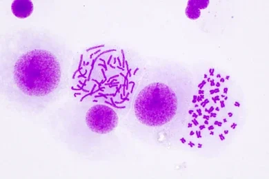
Karyotyping hematological malignancy
Karyotyping Hematological Malignancy is a cytogenetic test that analyzes the number and structure of chromosomes in blood or bone marrow cells. It detects chromosomal abnormalities such as translocations, deletions, inversions, or duplications associated with various hematological malignancies (blood cancers). This information is crucial for diagnosing the specific type of blood cancer, assessing its prognosis, and guiding personalized treatment decisions.
Specimen Type:
HEPARIN BM
TAT:
5-6 Working days
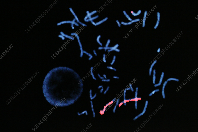
FISH leukemia ALL/AML
FISH Leukemia ALL/AML (Fluorescence In Situ Hybridization for Acute Lymphoblastic/Myeloid Leukemia) is a cytogenetic test that uses fluorescent probes to detect specific chromosomal abnormalities associated with acute leukemias. It identifies common translocations, deletions, and amplifications in leukemia cells, helping to diagnose the specific type of leukemia (ALL or AML), assess its prognosis, and guide personalized treatment decisions.
Specimen Type:
HEPARIN WB /BM
TAT:
5-6 Working days
FISH CLL
FISH CLL (Fluorescence In Situ Hybridization for Chronic Lymphocytic Leukemia) is a cytogenetic test that uses fluorescent probes to detect specific chromosomal abnormalities associated with chronic lymphocytic leukemia (CLL). It identifies common deletions in chromosomes 11q, 13q, 17p, and trisomy 12, which have prognostic significance and can guide treatment decisions for CLL patients.
Specimen Type:
HEPARIN WB /BM
TAT:
5-6 Working days

FISH MDS
FISH MDS (Fluorescence In Situ Hybridization for Myelodysplastic Syndromes) is a cytogenetic test that uses fluorescent probes to detect specific chromosomal abnormalities associated with myelodysplastic syndromes (MDS). It helps identify deletions, duplications, or rearrangements in chromosomes commonly affected in MDS, such as chromosomes 5, 7, 8, and 20. This information aids in diagnosing MDS, classifying its subtype, assessing prognosis, and guiding treatment decisions.
Specimen Type:
HEPARIN WB /BM
TAT:
5-6 Working days

FISH Myeloma
FISH Myeloma is a cytogenetic test that uses fluorescent probes to detect specific chromosomal abnormalities associated with multiple myeloma. It identifies common translocations, deletions, and amplifications in myeloma cells, which helps determine the disease’s risk category, prognosis, and potential response to treatment.
Specimen Type:
HEPARIN WB /BM
TAT:
5-6 Working days
FISH BCR ABL
FISH BCR-ABL1 (Fluorescence In Situ Hybridization for BCR-ABL1) is a cytogenetic test that uses fluorescent probes to detect the BCR-ABL1 fusion gene, a hallmark of chronic myeloid leukemia (CML) and some types of acute lymphoblastic leukemia (ALL). This fusion gene is formed due to a translocation between chromosomes 9 and 22, resulting in the Philadelphia chromosome. FISH BCR-ABL1 helps diagnose these leukemias, monitor treatment response, and detect minimal residual disease.
Specimen Type:
HEPARIN WB /BM
TAT:
3 Working days
FISH PML RARA
FISH PML-RARA (Fluorescence In Situ Hybridization for PML-RARA) is a cytogenetic test that uses fluorescent probes to detect the presence of the PML-RARA fusion gene, a hallmark of acute promyelocytic leukemia (APL). This test is crucial for diagnosing APL, as it identifies the specific chromosomal translocation t(15;17) that creates the fusion gene. It can also be used to monitor treatment response and detect minimal residual disease.
Specimen Type:
HEPARIN WB /BM
TAT:
3 Working days

FISH CMYC
FISH cMYC (Fluorescence In Situ Hybridization for MYC) is a cytogenetic test that uses fluorescent probes to detect abnormalities in the MYC gene, located on chromosome 8. This gene is involved in cell growth and proliferation, and its amplification or translocation can contribute to the development of various cancers, including Burkitt lymphoma and diffuse large B-cell lymphoma. FISH cMYC helps identify these abnormalities, aiding in diagnosis, prognosis, and treatment decisions.
Specimen Type:
HEPARIN WB /BM
TAT:
5-6 Working days

FISH double hit Lymphoma
FISH Double Hit Lymphoma is a cytogenetic test that uses fluorescent probes to identify rearrangements in the MYC gene along with BCL2 and/or BCL6 genes in lymphoma cells. The presence of these rearrangements (“double hit” or “triple hit”) is associated with a more aggressive form of lymphoma and helps guide treatment decisions and prognosis assessment.
Specimen Type:
Tissue biopsy / Paraffin Block
TAT:
5-6 Working days
